Glaucoma
Glaucoma refers to a group of diseases that cause damage to the optic nerve as a result of increased pressure within the eye, but can also be caused by a severe eye infection, injury, blocked blood vessels or inflammatory conditions of the eye.
There are two main types of glaucoma: Open-angle glaucoma and Angle-closure glaucoma. Open-angle glaucoma is the most common type of glaucoma and involves fluid in the eye not draining properly through the trabecular meshwork. Angle-closure glaucoma involves a sudden buildup of pressure in the eye and poor drainage because the angle between the iris and the cornea is too narrow.
Many patients do not experience any symptoms during the early stages of glaucoma, including no pain and no vision loss. This makes it difficult for many patients to know if they have the disease. But as glaucoma progresses, patients may experience a loss of peripheral or side vision, along with sudden eye pain, headache, blurred vision or the appearance of halos around lights.
nGoggle allows assessment of brain function using virtual reality – original video from Healio
Diagnosing Glaucoma
Since many patients do not experience any symptoms during the early stages of glaucoma, a diagnosis may only be made during a regular eye exam. It is important for patients, especially those with a higher risk of developing glaucoma, to have their eyes checked at least once a year.
Your doctor may perform several different eye exams in order to detect signs of glaucoma. Visual field testing is one of the most common tests performed, and involves creating a map to assess peripheral vision. Blind spots within the peripheral vision are often a warning sign for eye diseases like glaucoma.
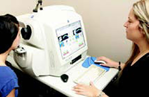
The Cirrus Optical Coherence Tomography (OCT) is one of the latest developments in diagnostic technology and is available at Prism Eye Institute. This testing system produces a highly detailed image of the structures and tissue within the eye to assess the thickness and health of the retina, macula, optic nerve and more, all without even touching the eye.
Diagnostic Technology
Eye on the latest in retinal diagnostic technology: Optical Coherence Tomography (OCT™)
This high-tech equipment allows physicians to clearly see the inside of the eye and treat problems before they progress. With this tool, we can see a highly detailed image of the eye structure and tissues. It especially allows us to see the back of the eye to better measure the thickness of the retina, macula, and optic nerve This technology helps us to accurately monitor diseases and conditions such as glaucoma, diabetic retinopathy, and uveitis.
- The instrument never touches the eye, so there is no discomfort.
- It provides a detailed analysis of all the layers of the retina in just minutes.
- For glaucoma, the Retinal Nerve Fibre Analysis identifies any glaucoma-related defects well before the patient would notice any symptoms.